Development of an open-source measurement system to assess the areal bone mineral density of the proximal femur from clinical CT images
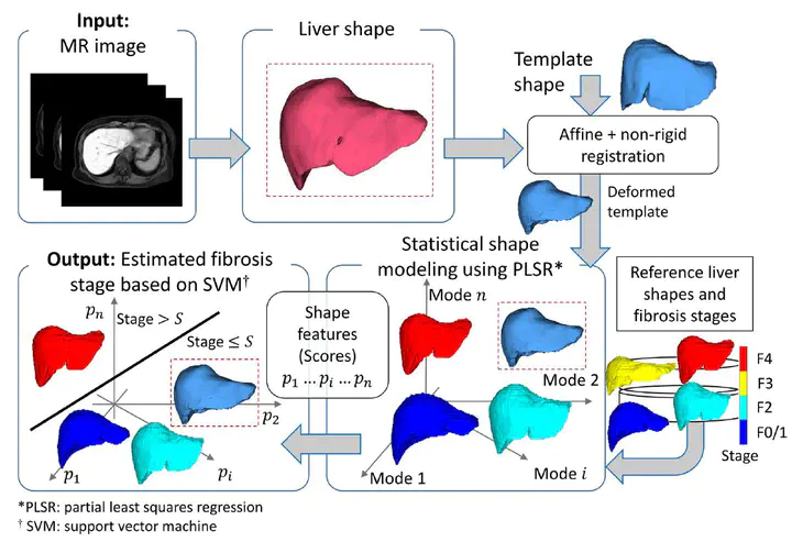
 Overall scheme for modelling the shape variations associated with liver fibrosis stage and predicting it fromMR images by using PLSR-based shape features (scores)
Abstract
Commercial software is generally needed to measure the areal bone mineral density (aBMD) of the proximal femur from clinical computed tomography (CT) images. This study developed and verified an open-source reproducible system to quantify CT-aBMD to screen osteoporosis using clinical CT images.
Type
Publication
Archives of Osteoporosis